AP C1 AND C2 - ODONTOID
Open Mouth Projection • Evaluation of odontoid process and C1-C2 vertebrae
CRITICAL WARNING - CERVICAL TRAUMA
DO NOT MOBILIZE NECK WITHOUT MEDICAL CONSULTATION
If cervical trauma is suspected:
- Do not mobilize neck without consulting physician
- Physician must examine patient after cervical lateral radiograph
- Follow cervical immobilization protocols
- Prioritize patient safety over image acquisition
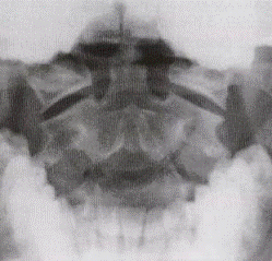
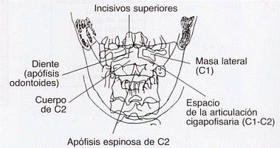
Demonstrated Pathology
Main purpose: Evaluate integrity of axis (C2) odontoid process
Key indication: Suspected fracture by flexion-extension mechanism
Exposure Factors
High kV: Necessary to penetrate skull base and visualize odontoid
Visible Anatomical Structures
Main focus: Clear visualization of odontoid between occiput and maxilla
Cassette Size
Orientation: Longitudinal
Justification: Sufficient to cover C1-C2 vertebrae and adjacent structures
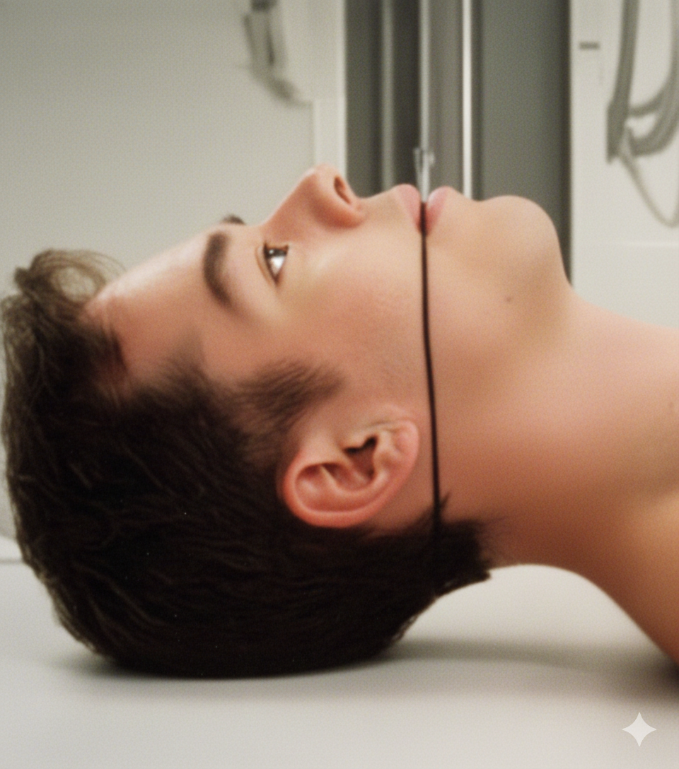
Patient Positioning
CRITICAL OPEN MOUTH POSITION
Mouth must be completely open to:
- Project upper and lower teeth away from odontoid
- Avoid dental superimposition over C1-C2
- Allow clear odontoid visualization
- Achieve incisors-skull base line perpendicularity
Verification: Patient must keep mouth open without moving jaw during exposure
Central Ray Point
Location: Directed to center of open mouth
Angulation: Perpendicular to table
Objective: Pass through open mouth to project odontoid free of superimpositions
Optimal Image Characteristics
Odontoid
Clearly visible without superimposition
Separated Teeth
Upper and lower incisors not superimposed
Skull Base
Projected above C1
Symmetry
C1 lateral masses symmetrical
Articular Spaces
Atlantoaxial joint visible
No Superimposition
Odontoid free of bony structures
Common Technical Challenges
Frequent problems in AP odontoid projection:
- Dental superimposition over odontoid from insufficient opening
- Head rotation causing asymmetry in C1 lateral masses
- Flexion/extension altering occiput-C1 relationship
- Poor patient cooperation to keep mouth open
- Degenerative arthritis hindering visualization
Solution: Verify incisors-skull base line perfectly perpendicular and mouth completely open
Patient Instructions
"Hold your breath during the exposure"
Complete sequence:
1. Place head against bucky
2. Open mouth completely
3. Maintain position without moving jaw
4. Hold your breath
5. Remain completely still
SPECIAL CONSIDERATIONS IN TRAUMA
In patients with suspected odontoid fracture:
- Do not force mouth opening if pain or limitation exists
- Consider alternative projection (Fuchs or Judet) if unable to open mouth
- Maintain cervical immobilization throughout procedure
- Constantly monitor patient for possible instability
Absolute priority: Cervical stability over image quality